The Regenerative Approach
BEFORE: There are some cases where people want to restore their missing teeth with implants but do not want to remove any teeth and bone. For these type of cases, a regenerative approach, instead of a reductive approach, can be performed. For the patient above, you can see the severe bone loss that has occurred in the upper arch of the mouth.
BEFORE: In a regenerative approach, hard and soft tissue (bone and gums, respectively) are regenerated within the area of the mouth in order to provide the support and proper health for the dental implants. Just for the patient’s upper left qaudrant, the patient will need to extract the unsalvageable teeth and undergo multiple procedures in order to restore the area properly with dental implants.
BEFORE: There is dimensional bone loss (bone reduction in height, width, and lenght) for the patient’s upper right quadrant. The x-ray shows only approximately 1.86mm of bone height, which is insufficient for supporting dental implants in this posterior area, especially where most of the masticational forces occur.
BEFORE: For the patient’s upper left side, the same condition exists as well. But for this region, there is significant bone loss at both the inside and outside of the sinus cavity. Furthermore, the bone loss in the anterior region within the quadrant will also be addressed as well. The white line illustrates the location of the desired crestal bone level.
AFTER: After the entire treatment has been completed for the upper arch, the patient is extremely grateful and relieved that no bone had to be reduced or “grinded down” as an end result for selecting a full-arch implant prosthesis at the upper arch. At the eight year recall (follow-up) as seen in the x-ray above, the patient is doing well and is problem-free.
AFTER: The procedures that were performed were bilateral external sinus elevations (aka sinus lifts) using BSM, vertical and horizontal bone augmentations via BBA, soft tissue augmentation, multiple implant placements, and final prostheses. The white arrows (within the posterior area of the mouth) and orange arrows (within the anterior area of the mouth) on the x-ray above indicate the new generated bone levels at both the alveolar crest and sinus cavity floor. The dental implants are now adequately supported by a sufficient volume of bone.
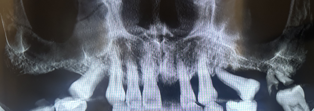
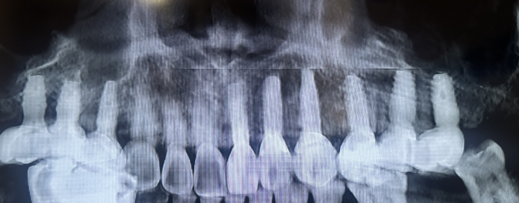
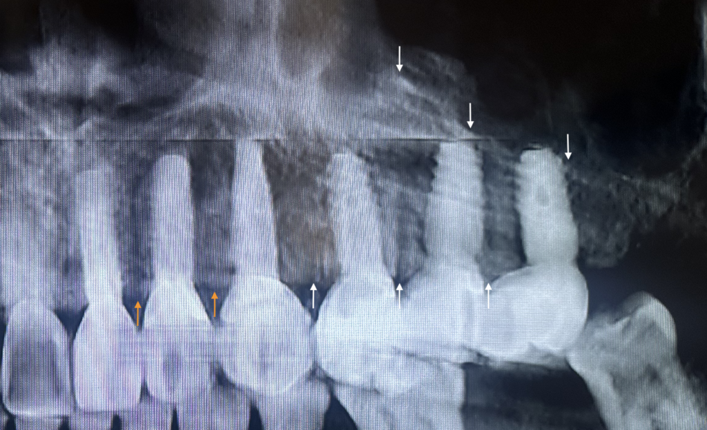
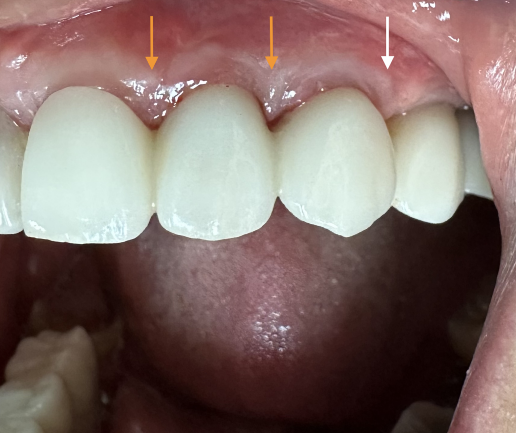
AFTER: Hence, by maintaining the appropriate alveolar crest and sinus floor bone levels, overall success for the implants can be achieved. Moreover, having the proper alveolar crest bone level in the anterior area (indicated by the orange arrows) can avoid and eliminate the “black triangles” (open spaces) that can be visible in between the teeth which are tremendously undesirable, particularly in the anterior esthetic zone. In the picture above, since the alveolar crestal bone level is at its desired height, the natural papilla of the gums can be seen to adequately occupy this specific area in between the teeth with no open space visible.