Biological Bone Augmentation (Bone Harvesting and Transplantation of Autogenous Bone)

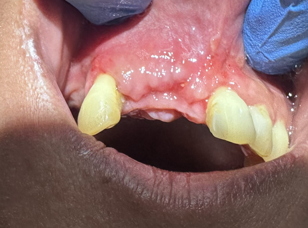
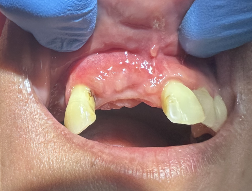
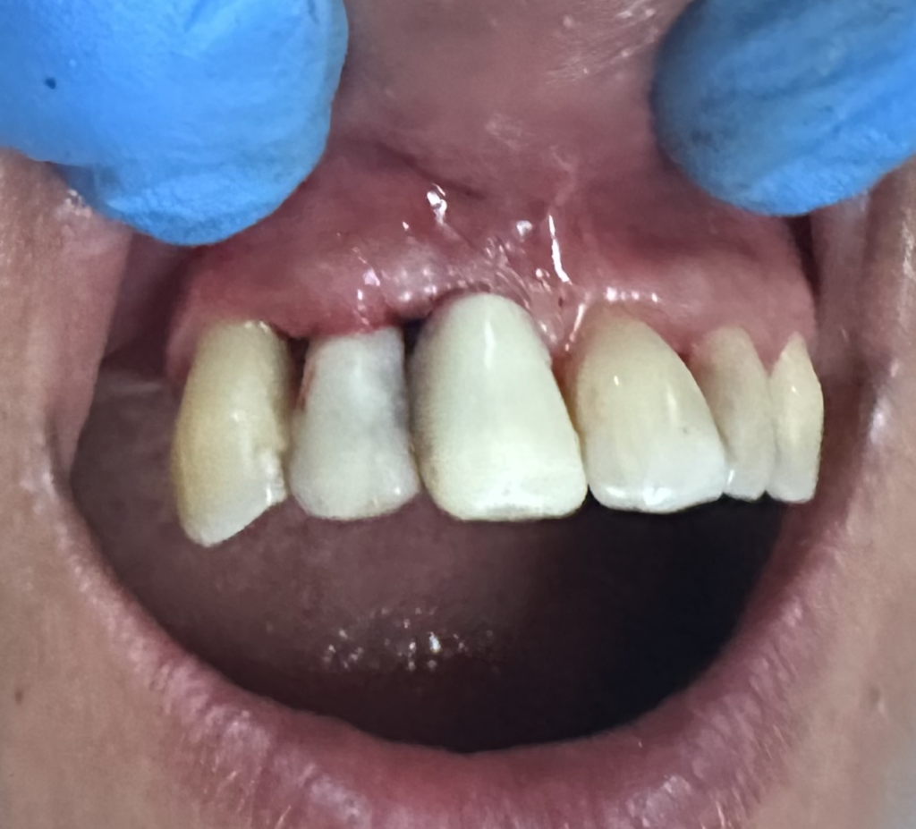
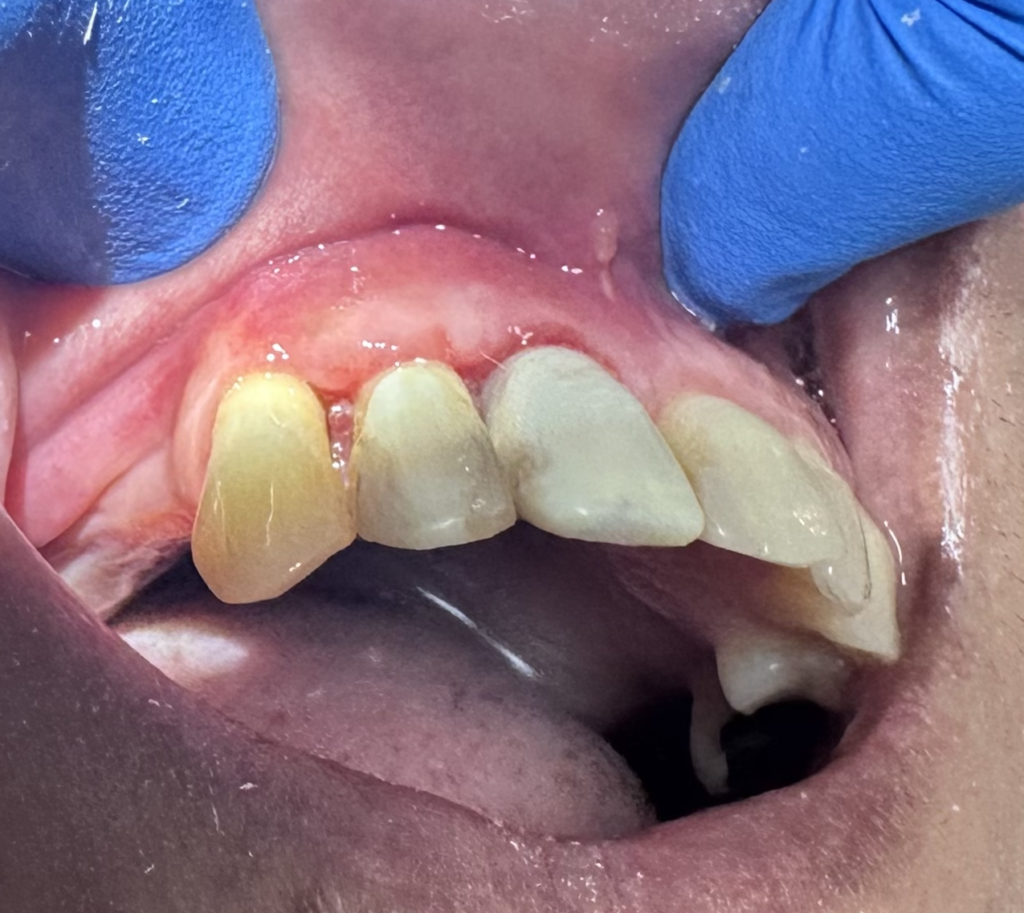
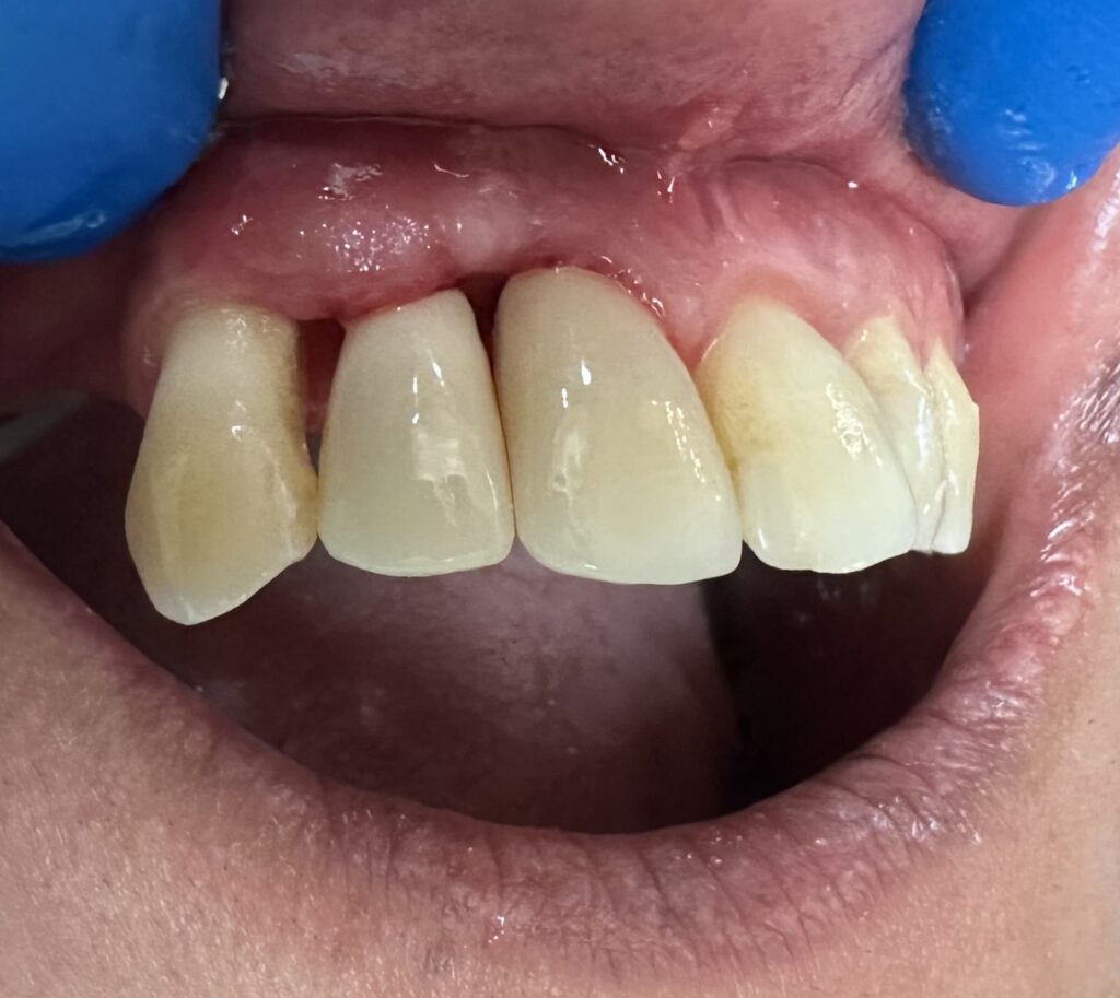
The patient has been in an accident in the past which resulted in the loss of two teeth. The patient has been wearing a removable partial denture (RPD) ever since and is now seeking to upgrade to a more permanent solution. 

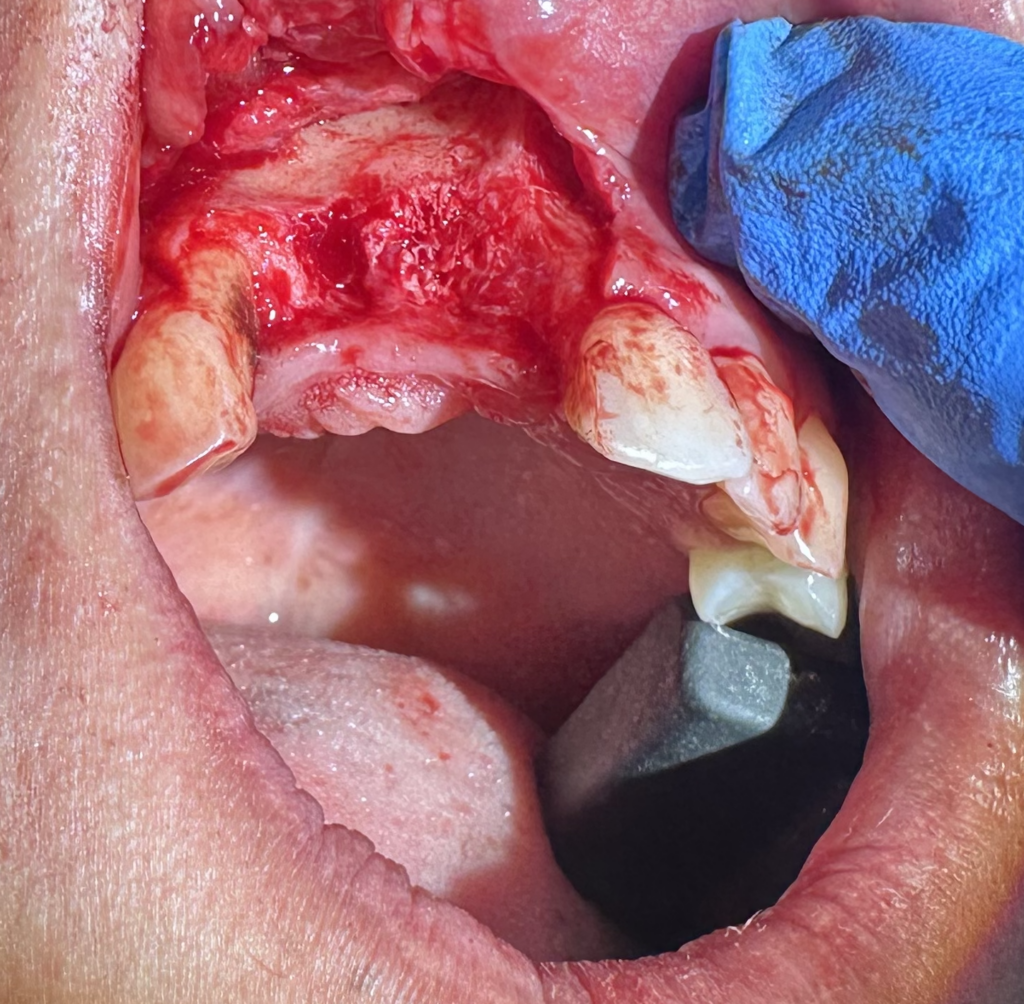
From the CBCT scan, it is shown that there is also a horizontal and vertical bony defect (also known as a “2-bony wall defect” since the front, back, and bottom walls are missing, leaving two walls on the sides) at the area of the missing teeth. From within the CBCT’s virtual planning software, a “virtual implant” is pre-planned and placed at the desired location of the site.
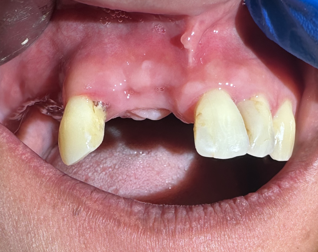
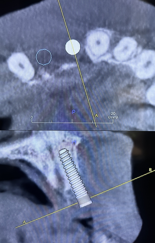
This area of missing bone will need to be “rebuilt” in order for implants to be placed. 

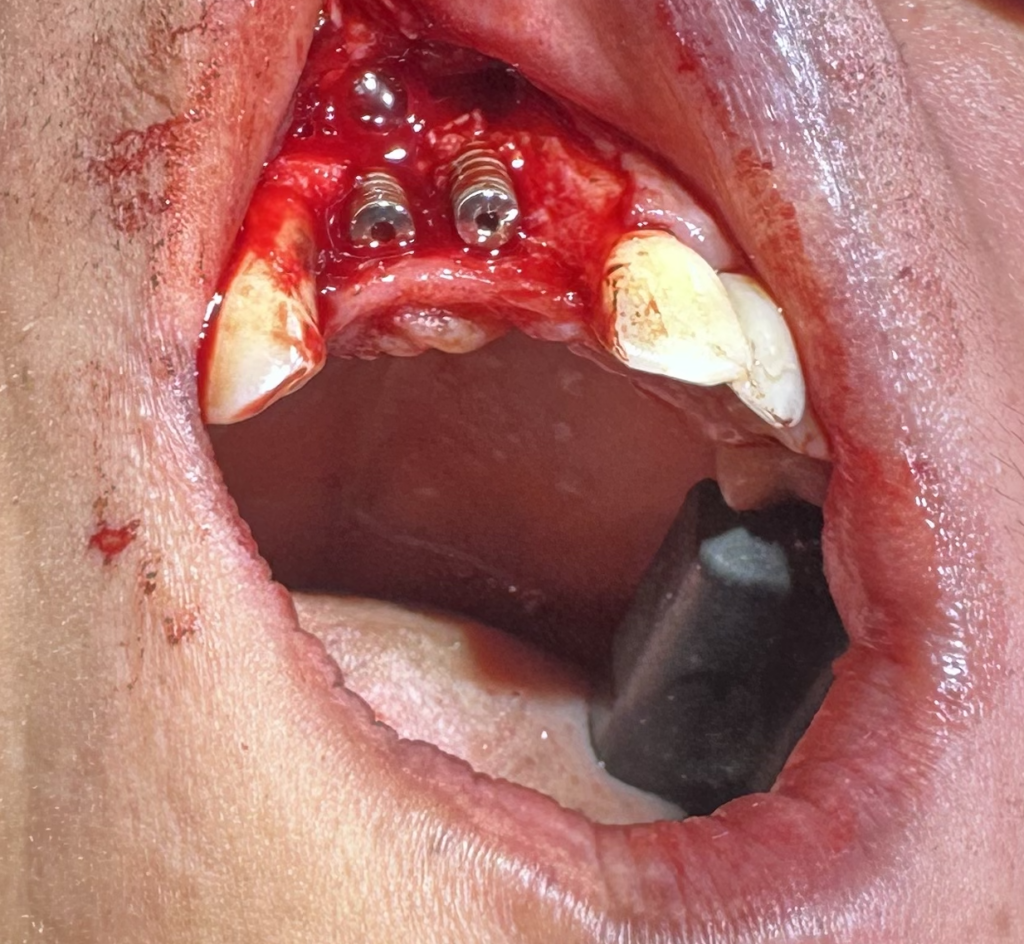
Two implants are placed freehandedly at the desired position.

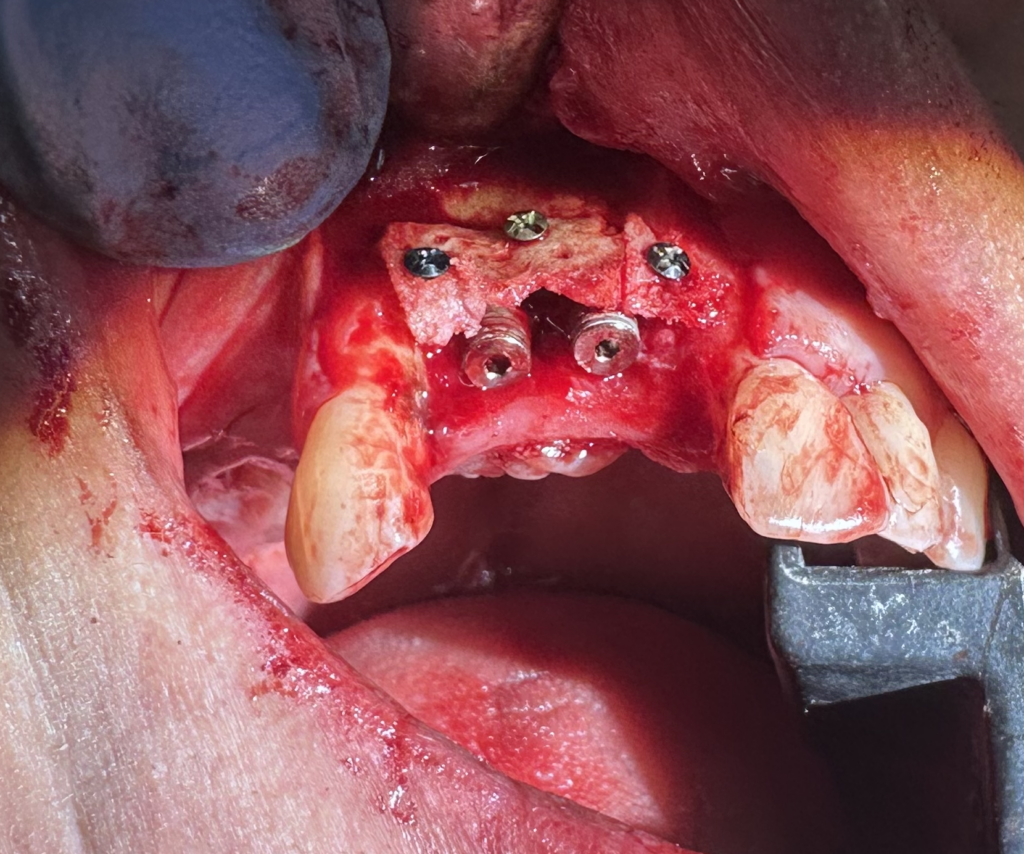
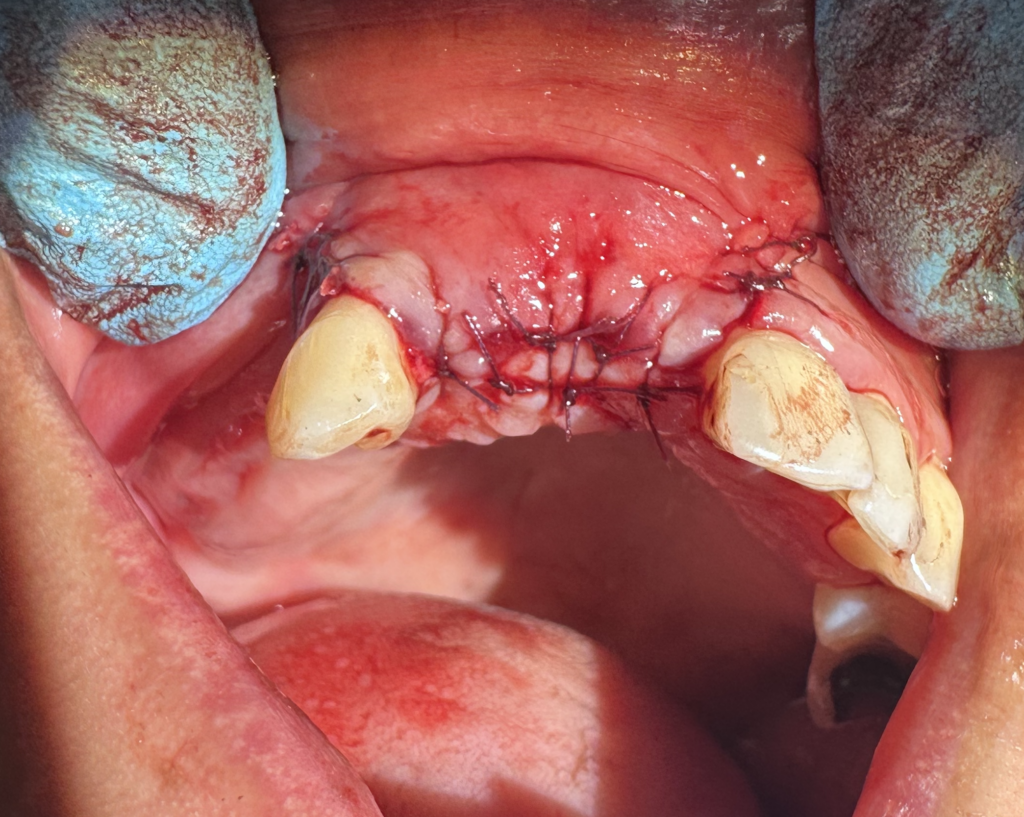
Autogenous bone (100%) onlay blocks are harvested from inside of the mouth and “bone shells” are created from them. The “bone shells” are then cut into smaller sections and made to fit and follow the curvature of the bone. They are used as wall barriers and help to contain bone particles that are made to a smaller size.

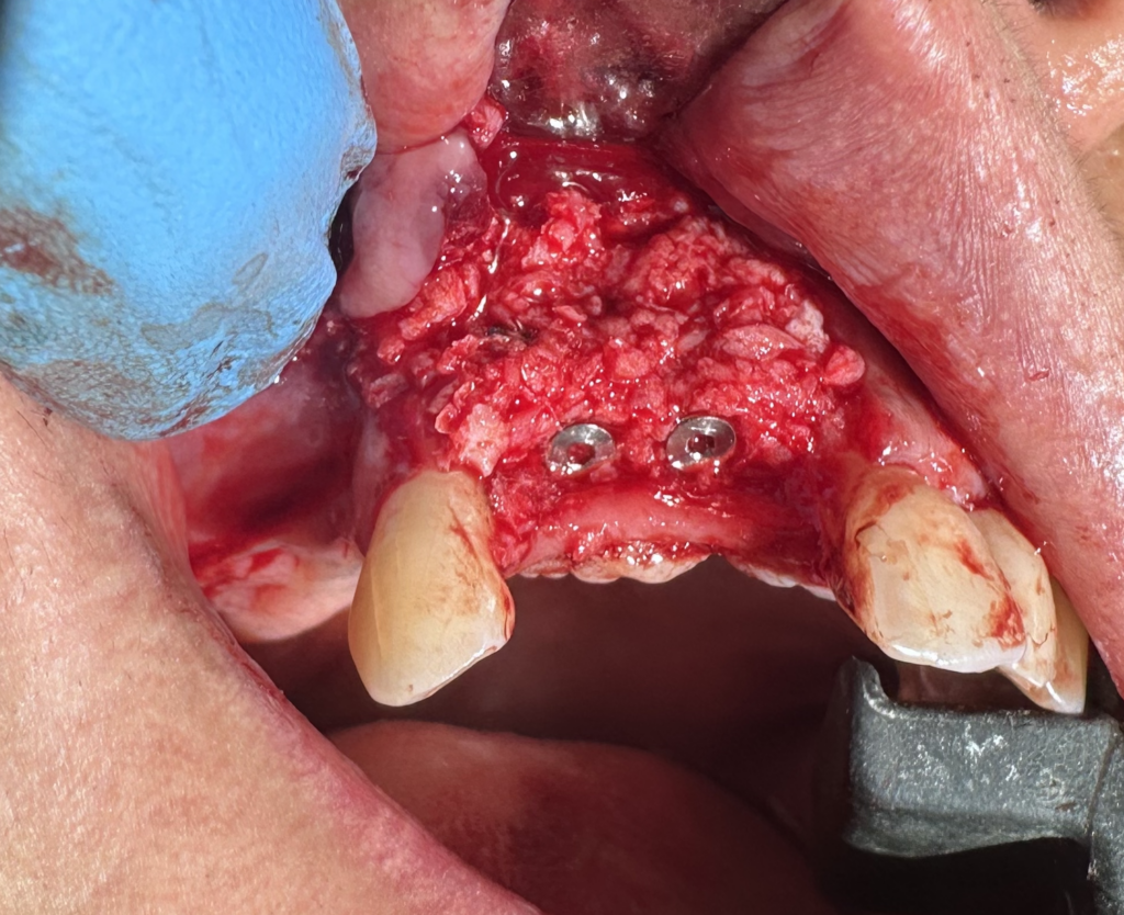
Autogenous (100%) bone chip and particulate particles (of sizes 250 microns – 1 mm and at 1 – 2mm, respectively), that are also harvested from inside of the mouth, are packed into the spaces within the “bone shells”.  Excess bone particles are added around the implant sites.

Tension-free primary wound closure is applied to the area to prevent incision line opening (ILO) from occurring. The implant site will then need to heal undisturbed and uneventfully for a desiginated period of time.

At the two-week follow-up appointment, all of the sutures are removed. It is re-entered at a later time for the next stage of treatment leading to the final fabrication of the implant prosthesis.

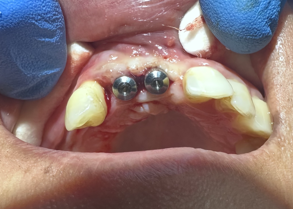
After four months of healing, the implants are prepared to be uncovered (exposed).

The implants are uncovered and permucosal extensions (PMEs) are attached to expand the gingival tissue and act as placeholders.

After a brief period during the same appointment, the PMEs are removed and replaced with chairside-made provisional (temporary) crowns. These crowns are then continuously shaped and modified to help establish the desired soft tissue drape and architecture surrounding the implants during healing. Additionally, a connective tissue graft can be harvested to help achieve ideal tissue esthetic harmony.

After a few weeks of soft tissue healing, the provisional (temporary) crowns are removed to reassess the height of the gingival soft tissue at the implant sites after the healing period and