Dental Implant Revision

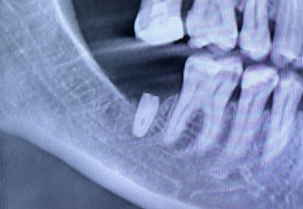
A patient is seeking a crown for an implant previously placed at another practice. However, the existing implant is critically short and narrow, which likely stems from an underlying lack of bone volume at the time of placement.

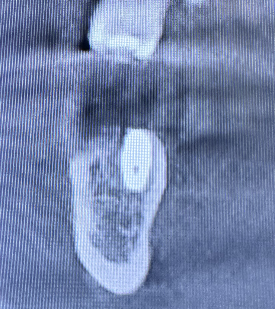
The cross-sectional CBCT view also reveals that the implant was placed in a non-ideal position (along the z-axis) on the alveolar ridge due to insufficient bone volume. Therefore, placing and utilizing a crown on the existing implant would cause it to fail prematurely.

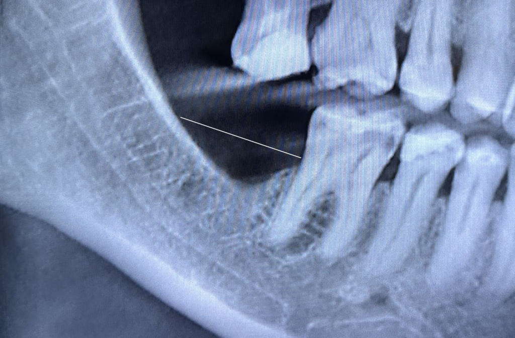
Consequently, the implant was removed, and a replacement was planned. The white line on the X-ray indicates the optimal bone height required for proper implant positioning, size selection, and crown-to-root ratio.

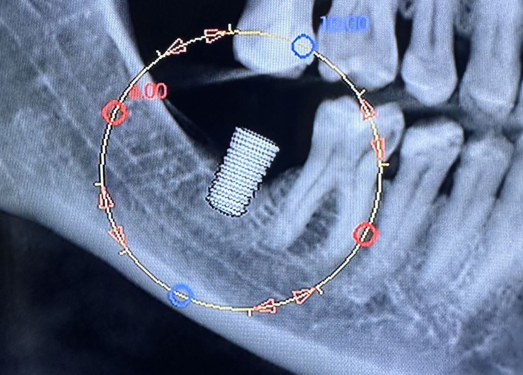
Using the CBCT’s built-in virtual planning software, a virtual implant of the intended size is placed at the desired location. Subsequently, new bone must grow to completely surround the implant.

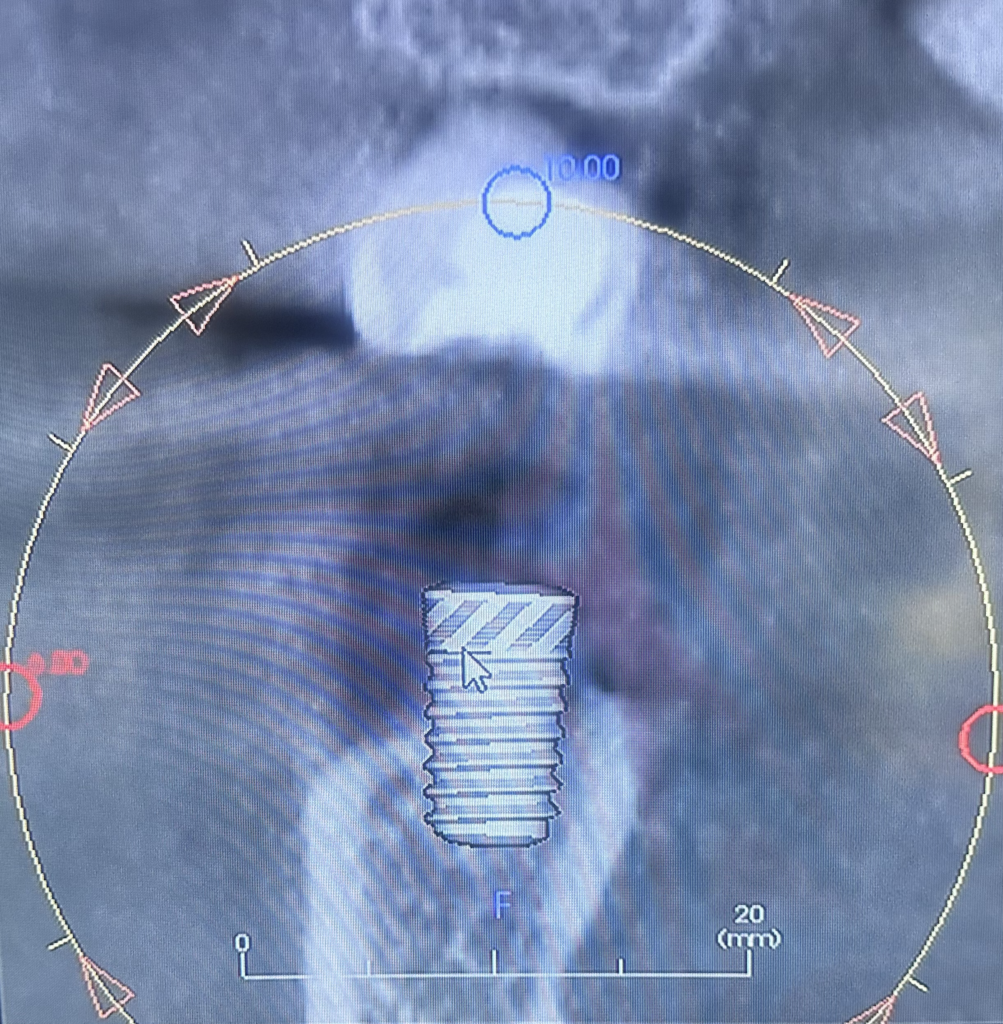
Moreover, the cross-sectional view of the CBCT scan indicates how much bone must be regenerated to fully encapsulate the virtual implant from all sides.

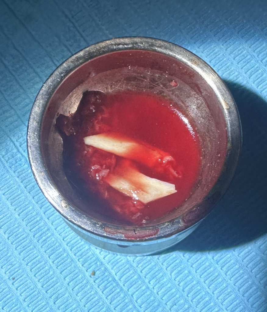
For this case, 100% autogenous bone graft is harvested from an intraoral donor site. Enough bone is obtained to correct the horizontal and vertical jaw bone defects and restore the volume required to support an implant at the site.

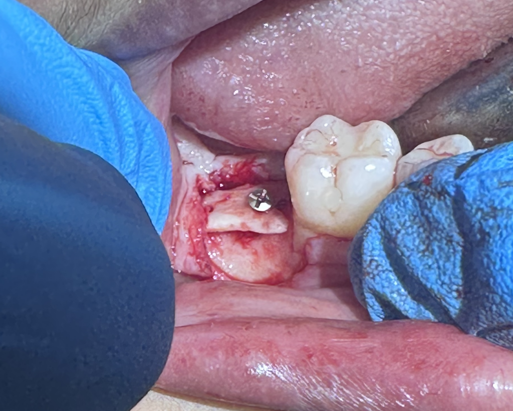
To access the recipient site, a tunnel flap is created to establish the upper border of the intended bony ridge height. Hence, a bone graft of a particular shape is harvested from the patient’s jawbone to serves as the upper wall, or ceiling, and to provide support.
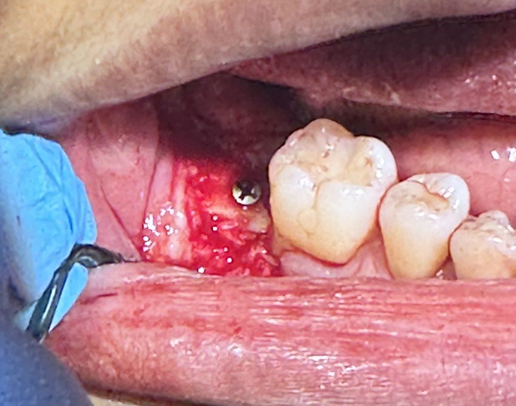
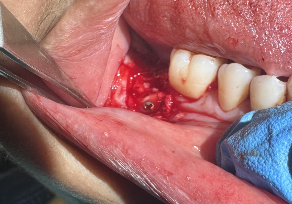
Next, autogenous bone chip particles are placed underneath the upper border to completely fill the gap between it and the native bone (alveolar ridge) from below.

A lateral (side) border was also harvested and added to act as an outer wall and aid in containing the autogenous bone chip particles confined to within the site.

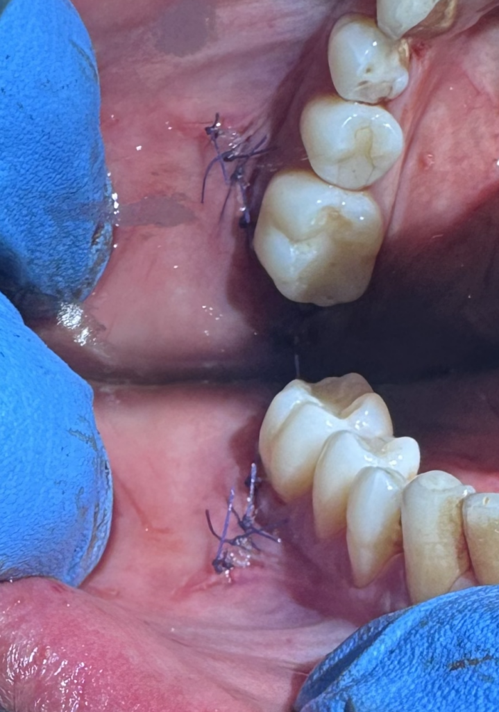
The flap is closed and without tension. Once the bone heals, the site is revisited to place the new implant.